Editorial standards
Image credits
Every image on this site is real photography or real microscopy. None is AI-generated. People are shown only from behind, in silhouette or at a distance, so no one is identifiable and no one appears to endorse us.
Kids shop illustrations and diagrams are hand-built SVG drawn by Everstack. Podcast covers are typographic; the official Spotify player loads only if you ask for it.
| Image | Description | Author | License | Source |
|---|---|---|---|---|
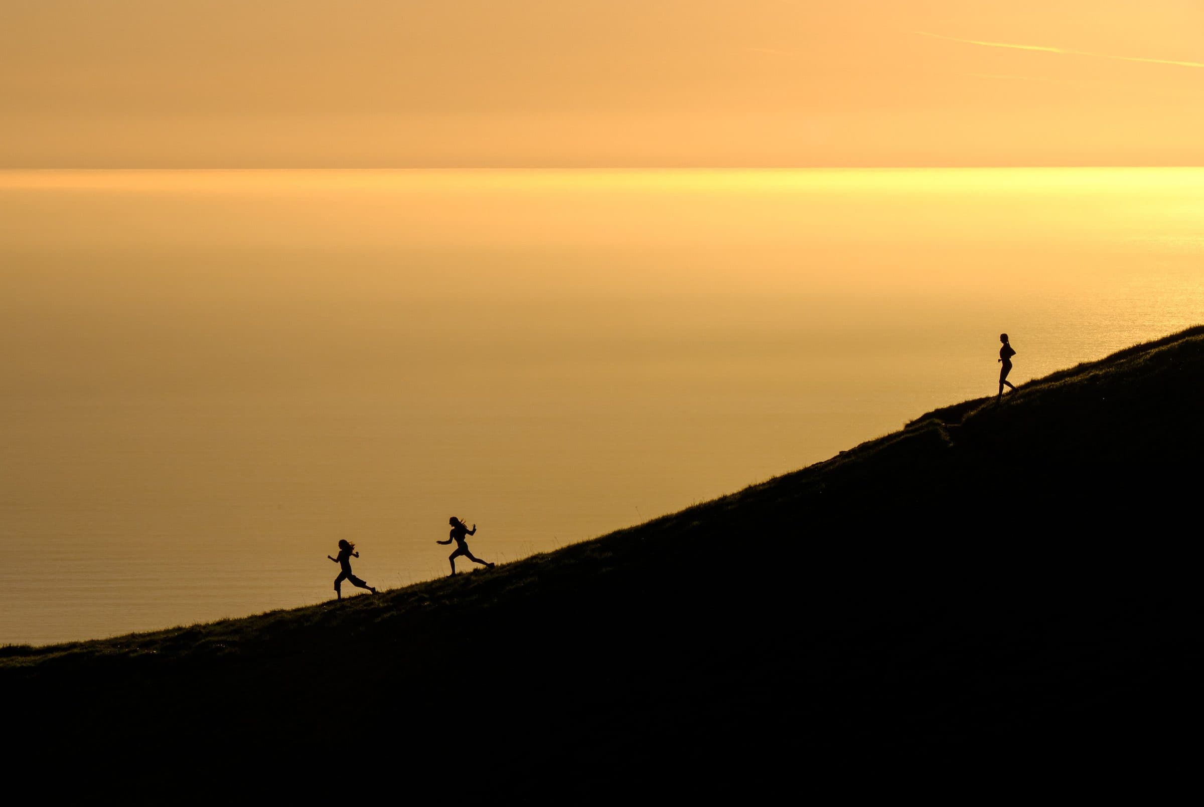 | Three runners in silhouette on a ridge at dusk Photograph | Todd Diemer | CC0 1.0 | Wikimedia Commons · Unsplash |
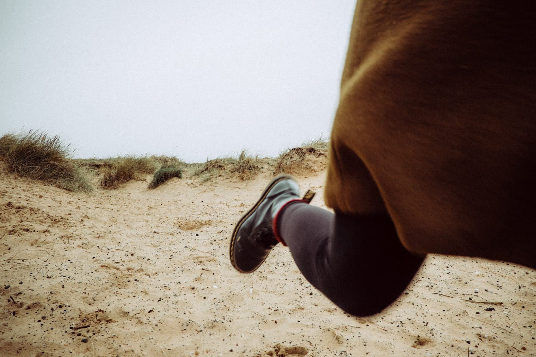 | A runner mid-stride on a sand dune, legs only Photograph | Mitchell Orr | CC0 1.0 | Wikimedia Commons · Unsplash |
 | A hiker seen from behind on a valley path Photograph | Matthew Kalapuch | CC0 1.0 | Wikimedia Commons · Unsplash |
 | A hiker from behind looking over a forested valley Photograph | Ales Krivec | CC0 1.0 | Wikimedia Commons · Unsplash |
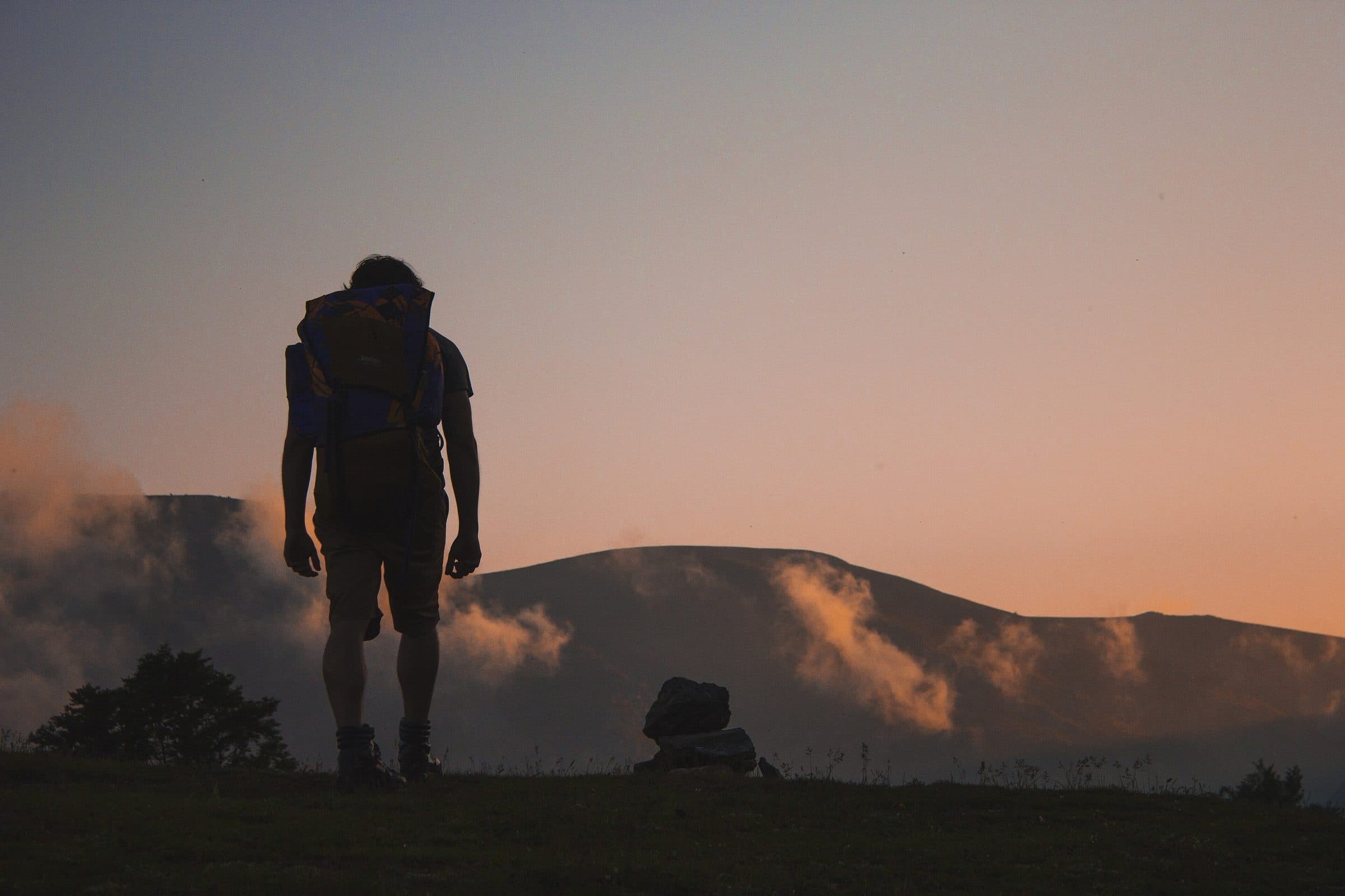 | A hiker in silhouette walking a ridge at dusk Photograph | Aneta Ivanova | CC0 1.0 | Wikimedia Commons · Unsplash |
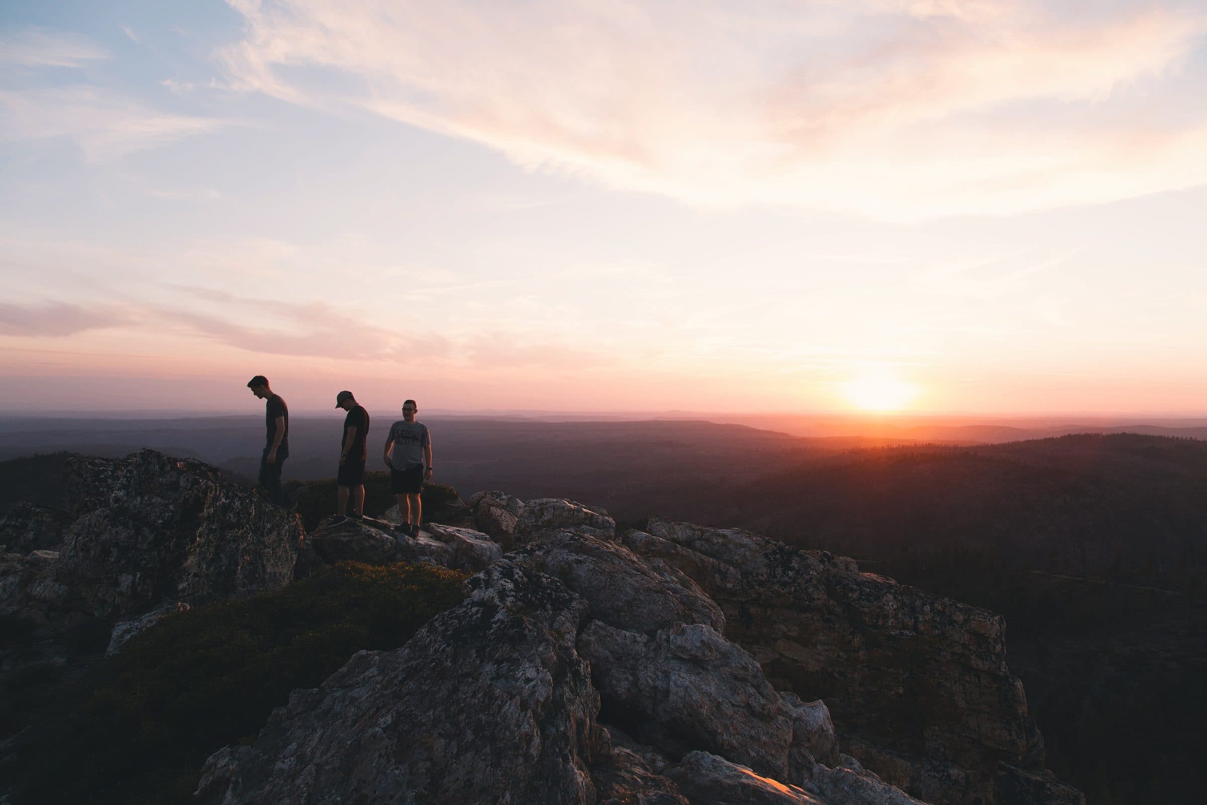 | Three people in silhouette watching a sunrise from rocks Photograph | Jordan McQueen | CC0 1.0 | Wikimedia Commons · Unsplash |
 | A person walking through a mountain meadow at golden hour Photograph | Cristina Gottardi | CC0 1.0 | Wikimedia Commons · Unsplash |
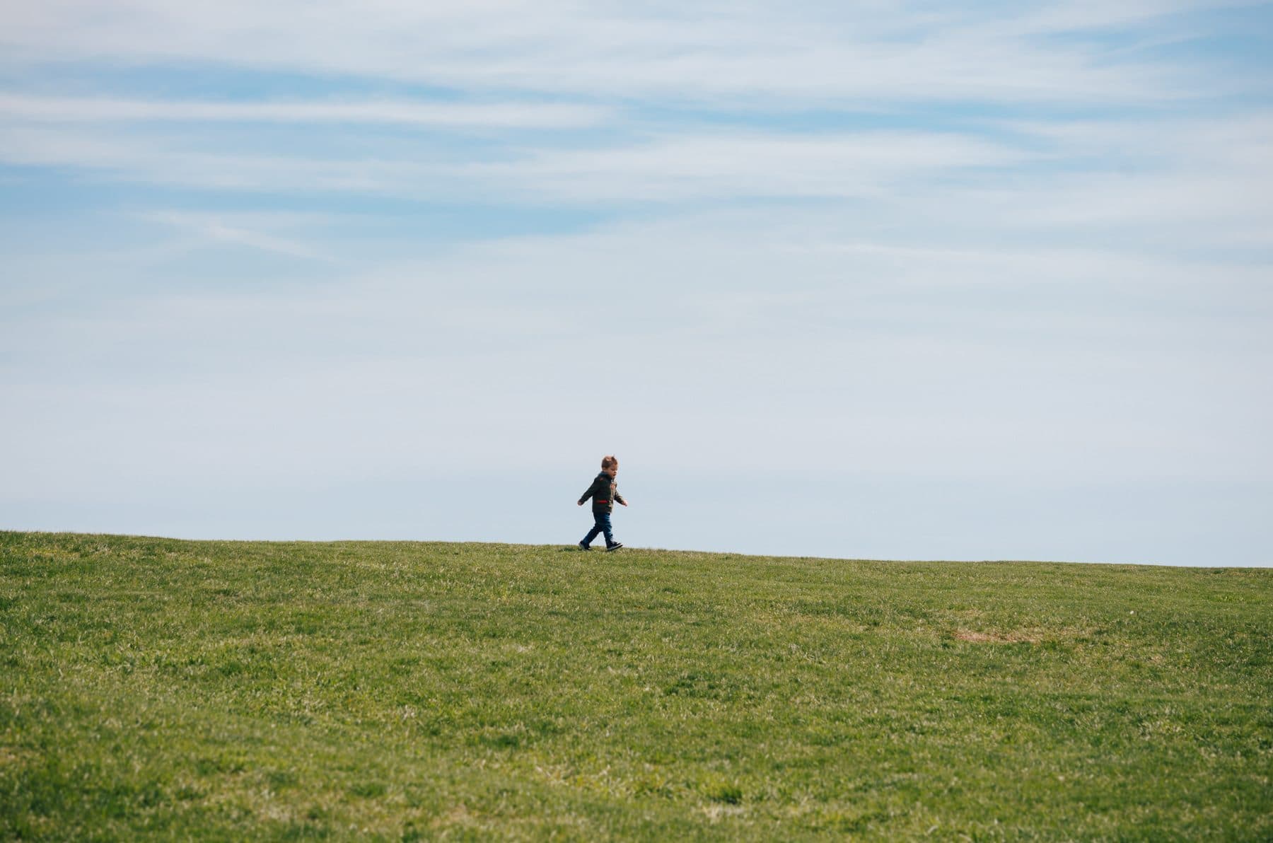 | A distant runner crossing a wide green field Photograph | Kelly Sikkema | CC0 1.0 | Wikimedia Commons · Unsplash |
 | A person in a balance pose in silhouette Photograph | Patrick Hendry | CC0 1.0 | Wikimedia Commons · Unsplash |
 | A cyclist climbing an empty mountain road Photograph | David Marcu | CC0 1.0 | Wikimedia Commons · Unsplash |
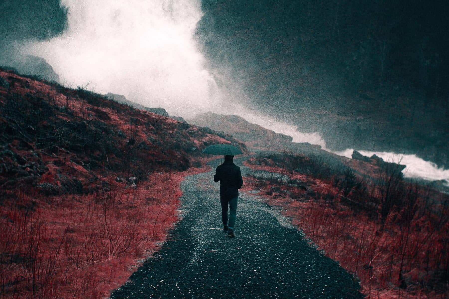 | A person walking a gravel road toward low cloud Photograph | Nick Scheerbart | CC0 1.0 | Wikimedia Commons · Unsplash |
 | A person from behind looking at a distant peak Photograph | Tim Bogdanov | CC0 1.0 | Wikimedia Commons · Unsplash |
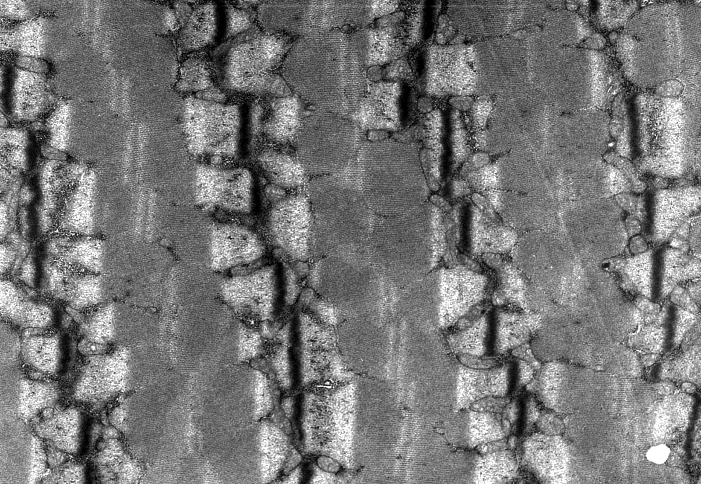 | Human skeletal muscle, electron micrograph: paired mitochondria sit beside each Z-line Microscopy | Louisa Howard, Dartmouth Electron Microscope Facility | Public domain | Wikimedia Commons |
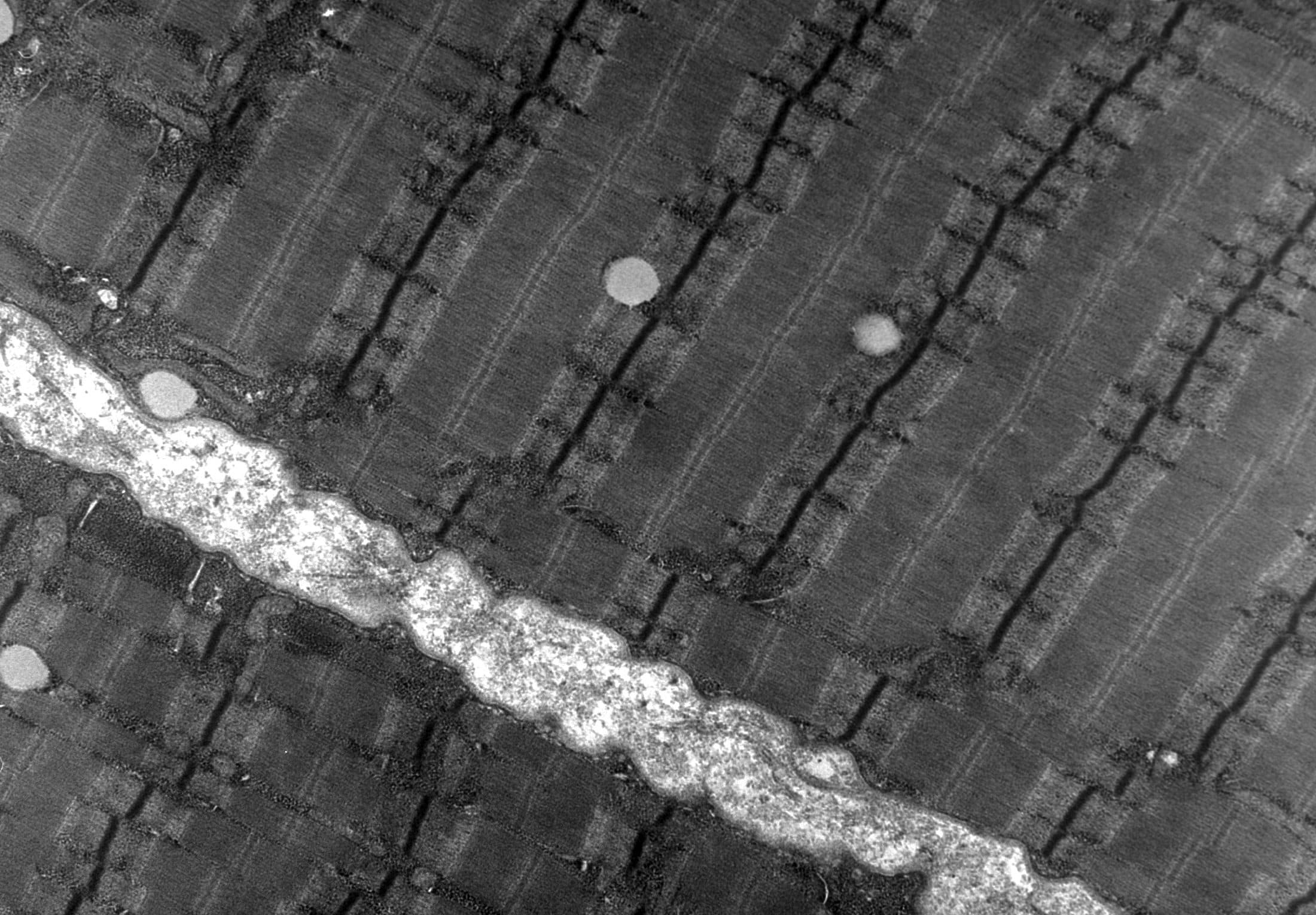 | Human skeletal muscle, electron micrograph: myofibrils and sarcomere bands Microscopy | Louisa Howard, Dartmouth Electron Microscope Facility | Public domain | Wikimedia Commons |
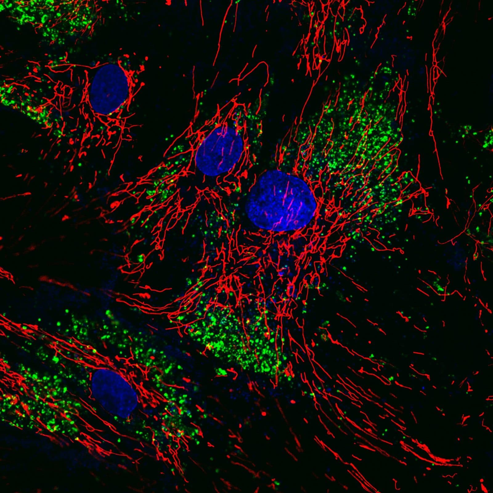 | Human fibroblasts, fluorescence: mitochondria in red, nuclei in blue, nanoparticles in green Microscopy | Matteo Battaglini | CC BY 4.0 | Wikimedia Commons |
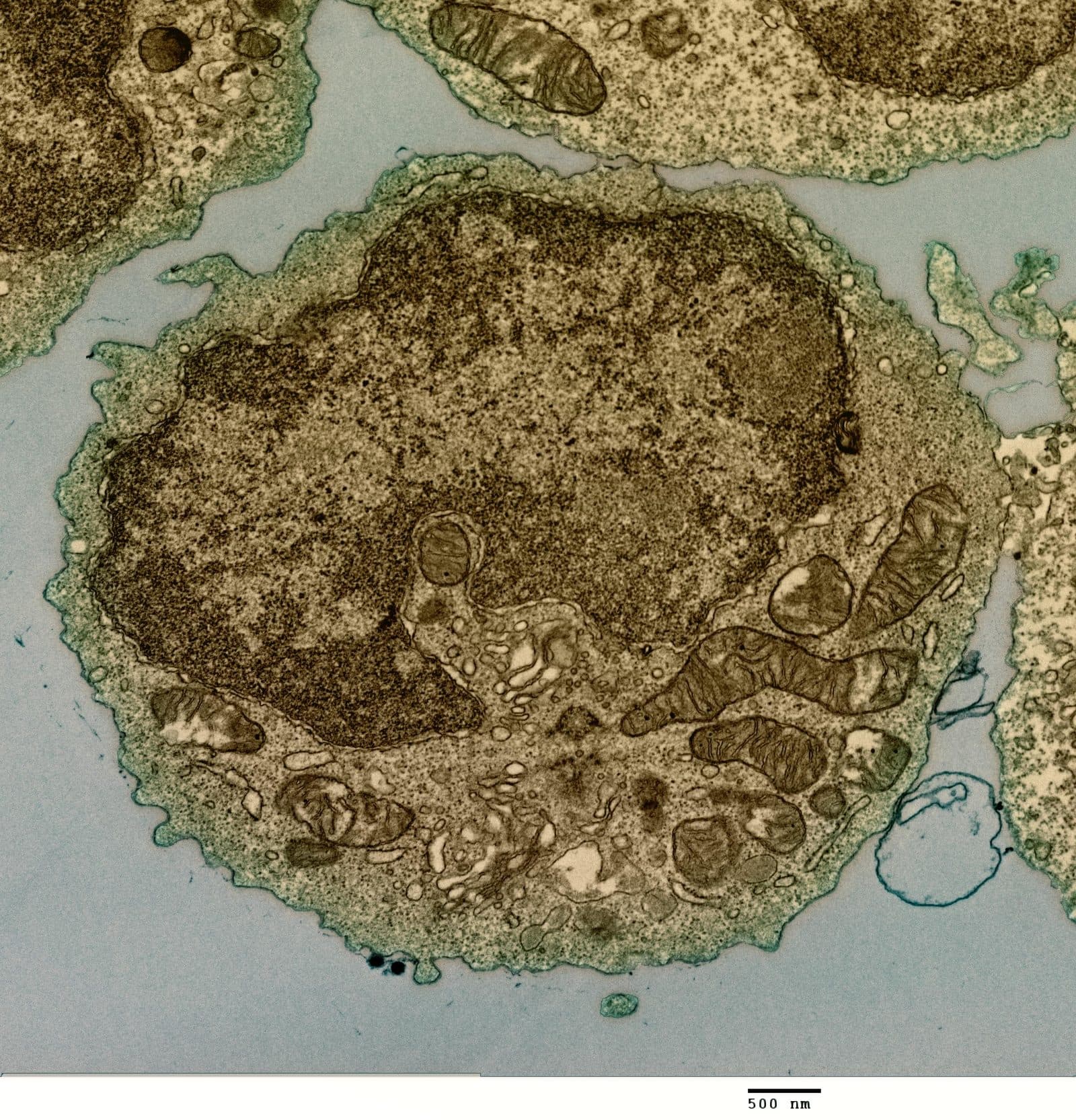 | A B cell from a human donor, transmission electron micrograph Microscopy | NIAID | CC BY 2.0 | Wikimedia Commons |
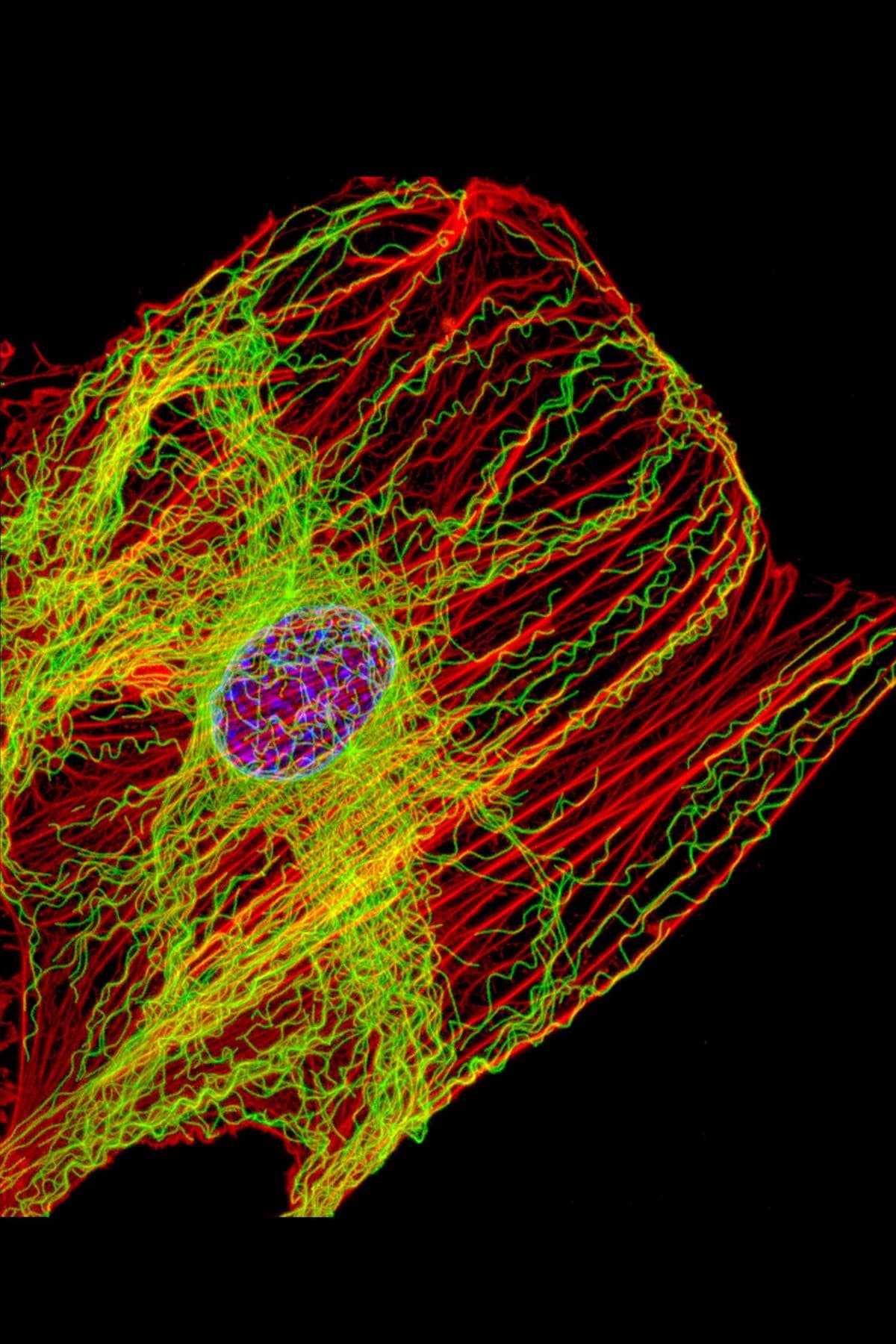 | A fibroblast, fluorescence: actin in red, microtubules in green Microscopy | James J. Faust and David G. Capco, NIGMS | Public domain | Wikimedia Commons |
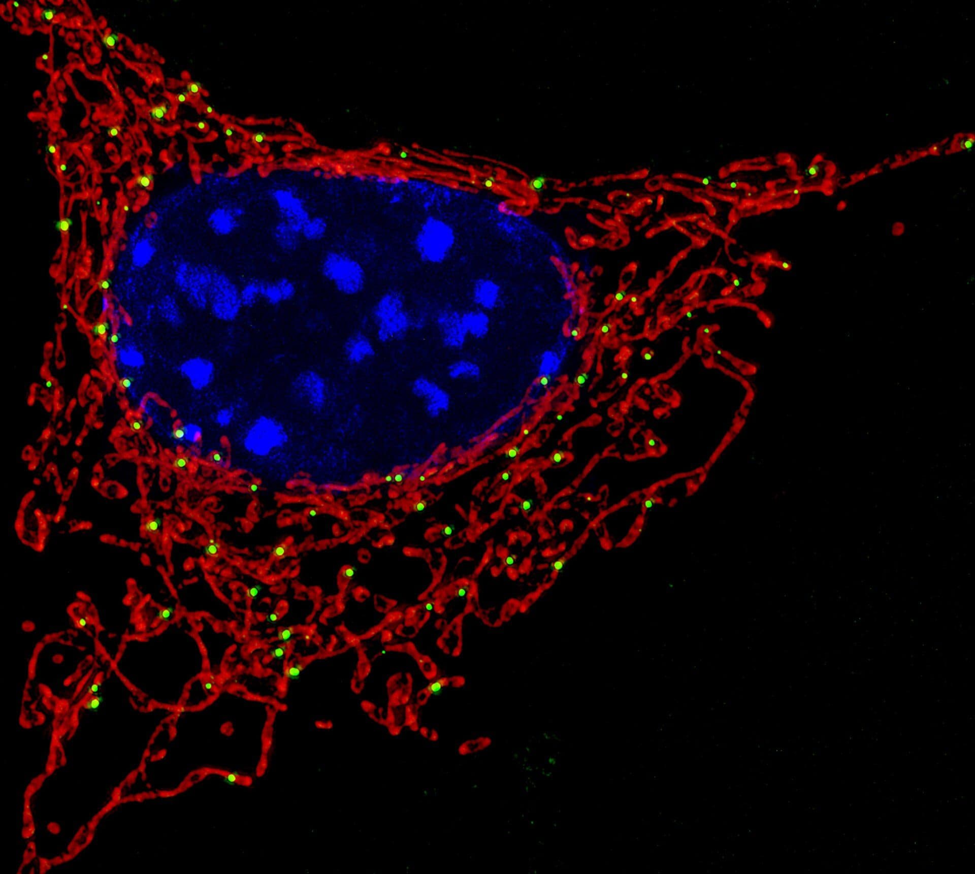 | A cell under fluorescence microscopy: the red network is its mitochondria, the blue is the nucleus Microscopy | NICHD, National Institutes of Health | Public domain | Wikimedia Commons |
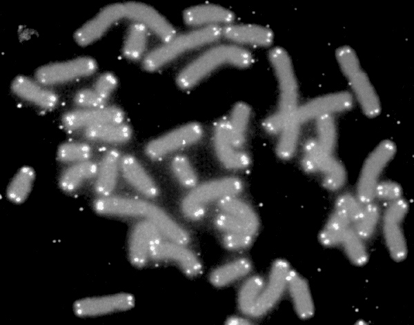 | Human chromosomes (grey) with telomeres lit at their tips (white) Microscopy | U.S. Department of Energy Human Genome Program | Public domain | Wikimedia Commons |
Licenses checked on Wikimedia Commons on 2026-10-15. Photos of people were originally published on Unsplash under CC0 1.0. Images are served optimized (AVIF or WebP) and are unaltered apart from cropping and scaling.